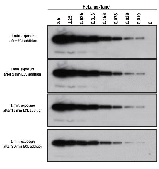
I have done a lot of collagen WB and they can be tricky but the nice thing is that the protein can take a pretty good beating and still come up. It will cut down on the protein you have, but should leave the insoluble matrix for your collection. If you really only need the ECM, try hypotonically lysing your cells from the plate first, then two washes of PBS and then the RIPA. Finally, you might be trying to run too much protein, so you're not getting good solubility. Switch to 4-25% gradient gels to help get your collagen in but keep your elastin from running off. This will really help loosen up your samples. Same thing for the tissue, although I'm not sure what "incubated the samples for 2 hours at 4 degrees" means? To your sample buffer add some DNAse. Change your denaturing step to 10% beta-Merceptoethanol for 10 minutes at 90-100. They really, really, want to stay crosslinked and depending on how long you are culturing your cells, the collagen might be in big fibrils. RIPA buffer usually has 1% SDS and that should be doing the trick so maybe it's your denaturing step. You might check if you can see a dark line at the top of the wells on your films, that could tell you that protein is getting stuck. HRP conjugated sheep anti-mouse, donkey anti-rabbit secondary antibodies, and the Enhanced Chemiluminescence (ECL) western blotting detection reagent were. Elastin is fairly small, but Collagen I and III are large and once everything is cross linked, it's like trying to get a gum ball to run into your gel. So the three things I can think of (other than the antibodies but I'm assuming they are previously tested for WB) are: 1) Your denaturation step , 2) Your proteins are stuck at the top of the well, I've seen this with WB for insoluble ECM, 3) Your ECM is still largely stuck on the plate. Well you're right to think that they might need another step. Something must be in the protein extract that inhibits the HRP-ECL select reaction, what could it be? This happens even if I use a different primary antibody and try to detect a different protein, the signal is still absent in this lane when using ECL select. When I develop this same blot with an Alkaline Phosphate conjugated antibody with BCIP and NBT I can see the band of interest. For that sample the lane is completely white, not even background signal is present. When I develop the blot with ECL select and image the membrane I can see my protein of interest in all of the samples, including my protein standard, except the cow manure sample.

Enhanced chemiluminescent (ECL) detection is based on antibodies conjugated to.

I develop the membrane with a typical Western blot protocol with a HRP conjugated secondary antibody. Choose the ECL Western blotting detection reagent that best suits your needs. The protein extract is run on an SDS-PAGE gel followed by a transfer onto PVDF membrane. The protein is collected by ammonium acetate precipitation. I have performed a SDS-phenol protein extraction on multiple samples including soil and cow manure samples.


 0 kommentar(er)
